|
|
|
Comentarios
|
|
|
El 10/10/2004 16:02, Antonio Félix Conde dijo:
Primary bronchioloalveolar carcinoma of the lung versus metastatic adenocarcinoma to the lung with bronchioloalveolar pattern. The presence of nuclear grooves favours the former possibility. Nevertheless, special techniques are required to make the differential diagnosis. Greetings, Antonio F. Conde Ibiza Spain
El 10/10/2004 21:18, David Cubero dijo:
Adenocarcinoma con patron de crecimiento bronquioloalveolar. Se necesitan los resultados de la IHQ y una imagen, aunque sea citológica del riñón para precisar. Si somos unicistas metástais de la mama. Saludos David
El 11/10/2004 0:19, Sandro Casavilca dijo:
La vascularización y fibrosis me sugieren la posibilidad de un hemangioma esclerosante con patron adenoide, a descartar carcinoma bronquiolo alveolar. Sandro Casavilca Hospital Central de la Fuerza Aérea. Lima - Perú.
El 11/10/2004 6:38, Giorgio Gherardi dijo:
Ciao a tutti The peripheral and subpleural location, as well as its architecture, the presence of apparently two types of cells lining the spaces (round and couboidal), and the sclerotic stroma also in my opinion suggest the so called pulmonary sclerosing hemangioma. The TTF-1+ / EMA+ / panCK- profile of the round cells would be of help to confirm this diagnosis.
El 11/10/2004 7:39, Ricardo Drut dijo:
"Tumorcito" pulmonar (pulmonary tumorlet; proliferación neuroendocrina). Ricardo Drut.
El 11/10/2004 8:06, César Ramírez dijo:
Hemangioma esclerosante. Saludos Complejo Hospitalario de Jaén España
El 11/10/2004 9:03, Bayardo Flores dijo:
La lesión aparentemente es única, sin embargo, me parece que puede corresponder a una Hiperplasia Adenomatosa Atípica (Adenoma Broquioloalveolar), aunque si en la última foto las atipias parecen más graves, por lo que me es difícil afirmar que no sea ya un Adenocarcinoma. Cordiales saludos a todos. Bayardo Flores Locarno, Suiza
El 11/10/2004 15:15, Larry Chen dijo:
El 11/10/2004 21:44, Leopoldo E. Santamaria dijo:
Hemangioma esclerosante..
El 12/10/2004 10:26, Maria Laura Fibbi dijo:
Io penso che si debbano prendere in considerazione almeno 2 ipotesi da verificare con immunoistochimica: 1)emangioma sclerosante 2)carcinoma bronchiolo-alveolare Dr. Maria Laura Fibbi
El 12/10/2004 14:34, Dan Pankowsky dijo:
Good morning colleagues! I hope your day is as nice is mine is here. I think this is most likely a primary bronchioloalveolar carcinoma based on its size and location (small peripheral lesion). I would normally sign out a case like this without immunohistochemistry, but with the history of a previous breast cancer and a concurrent kidney tumor, I would do TTF-1, Ck-7, GCDPF-15, to help. Bronchioloalveolar carcinomas should be strongly TTF-1 positive. Breast and lung can by CK-7 positive but it might help rule out renal cell carcinoma. GCDPF-15 will only help if it is positive.
El 12/10/2004 20:04, Vijay Singh dijo:
From the site, pattern and cells, I would think of Bronchoalveolar carcinoma.
El 13/10/2004 8:57, emilio mayayo dijo:
Hola a tod@s. En el macro-micro parece que son dos los nódulos. ¿alguien se apunta a Minute Pulmonary Meningothelial-like Nodules (pequeños nódulos meningoteliomatosos pulmonares)?. Aunque me sobra el patrón vascular y me faltan técnicas. Seguimos en el foro. Enhorabuena por la aceptación y cada vez más participación internacional. Emilio
El 13/10/2004 9:25, Nidhi Bhatt dijo:
I would go in favour of a bronchioloalveolar carcinoma of lung. However, I would like to see slides from the kidney tumor and previous breast Ca to rule out the possiblity of a metastic adenocarcinoma from either site ( papillary ca of kidney or breast)
El 13/10/2004 10:00, Dr S Jivaji dijo:
Greetings forum members, I think this is a bronchoalveolar carcinoma. However in view of the history I would want to review the previous tumor slides from the breast and also the kidney
El 13/10/2004 13:53, Túlio Geraldo de Souza e Souza dijo:
. Caros unineters, colocamos a sua disposição, mais duas imagens do caso, para facilitar, diagnósticos diferenciais. ~~~~~~~~~~~~~~~~~~~~~~~~~~~~~~~~~~~~~~~~~~~~~~~~~~ Dear unineters... You can see now, two new imagens of the case, that can help you in the differencial diagnosis. ~~~~~~~~~~~~~~~~~~~~~~~~~~~~~~~~~~~~~~~~~~~~~~~~~~~~ Great for attencion!!! Túlio. .
El 13/10/2004 15:41, Bayardo Flores dijo:
Las últimas fotos me inducen a retirar la propuesta de Adenoma vs. Carcinoma bronquioloalveolar. Ahora se aprecia con más claridad que la lesión está constituida por dos tipos celulares, el epitelio de revestimiento bronquiolar aparentemente no atípico e inmediatamente en posición subepitelial una población celular epitelioide con citoplasma mal definido y núcleos un tanto pleomórficos, con hendiduras (grooves), que recuerdan a células de Langerhans, aunque no pienso se trate de una histiocitosis. Con todo esto pienso que se podría agregar a las posibilidades diagnósticas ya señaladas por otros compañeros la de un Tumor Carcinoide Periférico. Seguimos errando, tal vez algún día nos toca aprender. Saludes Bayardo
El 14/10/2004 19:42, sonal kulkarni dijo:
From the cellular arrangement & site of the lesion I would prefer the diagnosis of primary bronchoalveolar carcinoma
El 15/10/2004 7:33, Maurizio Ferretti dijo:
Cari Colleghi, I guess the morphologic pattern allows to rule out a metastasis, both from kidney and breast carcinoma. The cubic cells we must idenify seems to growth beneath the layer of bronchiolar epithelium. Thus i think it could be a so called tumorlet. Obviously we need some diagnostic ancillary techniques (i.e. chromogranin) to reach a correct diagnosis. The second I fovour well differentiated bronchiolo alveolar carcinoma (it was my first diagnostic impression)and the third sclerosing hemangioma. Saluti a tutti Dott. Maurizio Ferretti Citopatologia Diagnostica Ospedali Riuniti di Ancona Italy
El 15/10/2004 12:01, MAURIZIO SPINELLI dijo:
La mia prima idea è Pneumocitoma (cosiddetto Emangioma sclerosante). Saluti a tutti Maurizio Spinelli (Milano)
Hacer un comentario a este caso
|
|
|
|
|
|
|
Comentario del Autor
|
|
|
|
.
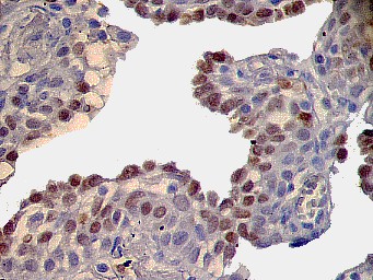
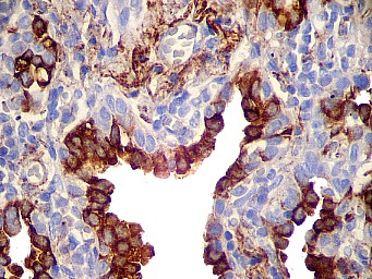
A neoplasia pulmonar apresenta dois tipos de células claramente distintas. As superficiais, sem as atipias esperadas no carcinoma bronquiolo alveolar. As células cúbicas, estratificadas mostram a morfologia típica das células redondas dos pneumocitomas. O primeiro tipo celular expressa TTF1, EMA, Spa, CK7 e AE1/AE3(Vide fotos adicionais). O segundo tipo expressa TTF1, CK7 e inesperadamente AE1/AE3 (Vide fotos adicionais). Este caso apresenta dois padrões: papilar e adenóide. O tumor renal é um clássico carcinoma de células claras e o tumor da mama um carcinoma ductal. Ambos sem semelhança com o tumor pulmonar.
The lung tumor has two cellular components clearly discernible. The superficial cells are not atypical as we expect in bronchioloalveolar carcinoma. The other cellular component is cuboidal and stratified, with the typical morphology of the round cells found in Pneumocytomas.
The first cellular type expresses TTF1, EMA, Spa, CK7 and AE1/AE3 (See new pictures). The second type expresses TTF1, CK7 and unexpected AE1/AE3 (See new pictures). This case show two patterns: papillary and adenoid. The kidneys tumor is a classic clear cell carcinoma and the breast tumor a ductal carcinoma. Both without similarity with the lung tumor.
.
|
|
|
|
TTF1
|
|
|
|
Surfactante (Spa)
| |
|
|
|
|
|
|
Bibliografía
|
|
|
. 1. Devouassoux-Shisheboran M, Hayashi T, Linnoila RI, Koss MN, Travis WD. A clinicopathologic study of 100 cases of pulmonary sclerosing hemangioma with immunohistochemical studies: TTF-1 is expressed in both round and surface cells, suggesting an origin from primitive respiratory epithelium. Am J Surg Pathol. 2000 Jul;24(7):906-916. 2. Lin D, Zou S, Lu N, Liu X, Wen P, Li L. Thyroid transcription factor-1 in the histogenesis of plumonary sclerosing hemangioma. Zhonghua Zhong Liu Za Zhi. 2002 Jul;24(4):384-387. 3. Chan AC, Chan JK. Pulmonary sclerosing hemangioma consistently expresses thyroid transcription factor-1 (TTF-1): a new clue to its histogenesis. Am J Surg Pathol. 2000 Nov;24(11):1531-1536. .
|
|
|
|